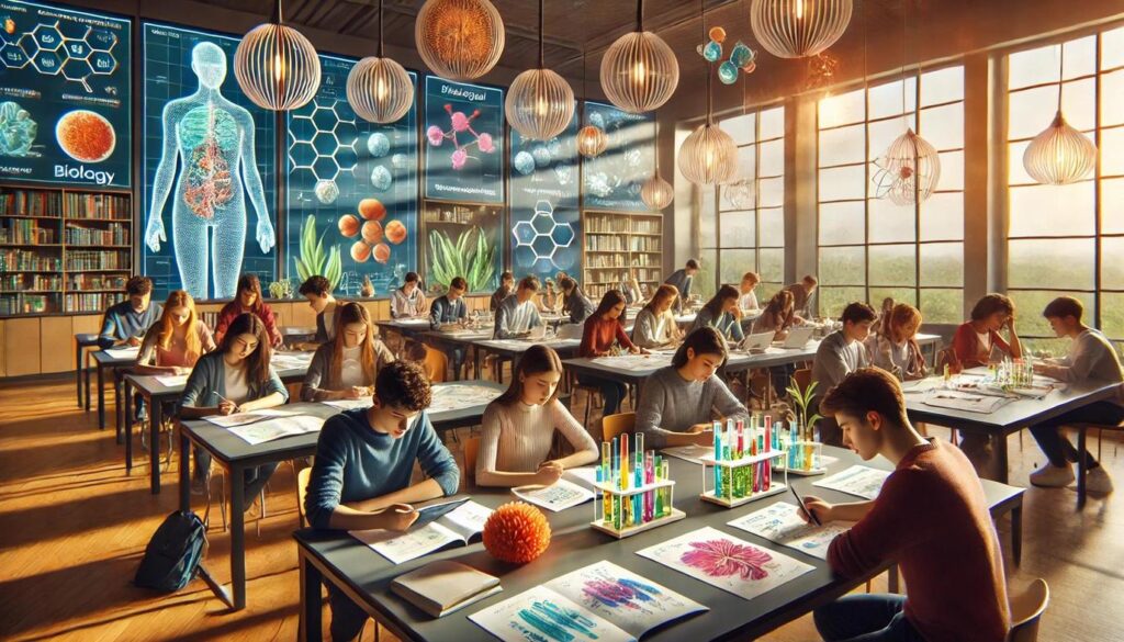
The best science classes engage students beyond textbooks. When learners get to physically inspect structures, manipulate real materials, and actively explore anatomy, they move from passive note‑takers to curious investigators. One of the most effective ways to embed this mode of learning is using preserved resources—such as high‑quality dissection specimens—that invite exploration, discussion and observational skill‑building.
Enhancing Engagement
Students often remember what they’ve done more than what they’ve read. A tactile lab where they hold, examine and ask “why is this shaped like that?” creates lasting impressions. Dissection brings the internal structure of organisms into tangible view and converts abstract concepts into observable reality.
Supporting Diverse Learning Styles
Visual learners benefit from seeing organ structures, kinesthetic learners from manipulating specimens, and verbal learners from describing their observations out loud. Hands‑on materials allow multiple learning modes to intersect. For many students, the shift from diagram to real object bridges the gap that textbooks can’t always fill.
Fostering Scientific Mindsets
Beyond the physical act of dissection lies the mindset of inquiry: students observe, hypothesise, test, record and reflect. These are the skills of scientists. Working with preserved models transforms a routine lab into an investigative experience.
Preparing for Advanced Study
For students planning to enter health sciences, veterinary fields or advanced biology, early exposure to realistic models of anatomy builds confidence. Recognising structures, understanding spatial relationships and practising manual precision become valuable foundations.
Choosing the Right Materials for Anatomy Labs
To make a real difference in learning, the equipment and resources must be thoughtfully selected. It’s not just about having specimens—it’s about having the right specimens and the right tools to make study effective.
Quality and Preservation Matter
Specimens that are clearly preserved, labelled and maintained allow students to focus on learning rather than struggling with artefact degradation. Vendors who specialise in anatomical specimens highlight the importance of proper preservation.
Well‑prepared materials reduce frustration and maximise instructional time.
Tool Kits and Supportive Equipment
Alongside the specimens you’ll need trays, pins, tweezers, scalpels, forceps and protective gear. A full lab kit ensures that all students can participate without delays or deficits in resources. A standard list might include:
- Dissecting pan and wax pad
- Scalpel handle with assorted blades
- Curved and straight forceps
- Dissecting pins and probes
- Safety goggles and gloves
Alignment with Curriculum
Select materials that map directly to your learning goals. If you’re teaching organ systems, comparative anatomy or invertebrate vs vertebrate structures, make sure the specimen set supports those themes clearly and precisely.
Ethical and Practical Considerations
Preserved specimens raise questions of sourcing, disposal and student perception. Clear procedures for handling, storage and respectful treatment help set a professional tone and reinforce the value of the lab.
Preparing Students for a Successful Dissection Experience
The lab environment matters. Preparation in setup, expectations and safety ensures smooth execution and maximises learning.
Pre‑Lab Orientation
Before the first cut is made, review safety protocols: wearing gloves, using tools responsibly, handling specimens respectfully and disposing of materials properly. Also clarify the learning objectives: what anatomical relationships are they exploring? What questions should they ask?
Setting the Stage for Observation
Encourage students to first look—examine external features, make sketches, note anomalies—before diving in. Observing surface anatomy, texture and colour builds observational muscles that strengthen during the dissection.
Team Roles and Rotation
Working in teams fosters collaboration. One student might handle documentation, another might manipulate tools, a third might identify structures. Rotating roles ensures no one is passive—and everyone gains skill.
Safety and Respectful Handling
Remind students that these are real biological specimens. The tone is science, not spectacle. Proper disposal—or storing for future reuse—shows respect and models professional lab behaviour.
Debrief and Reflection
After the active work is done, set aside time for students to discuss: What surprised you? What did you expect? How does what you saw match diagrams or textbooks? This reflection builds deeper understanding.
Suggested Pre‑Lab Checklist
- Review tool names and functions
- Discuss the organism’s external structure and likely internal layout
- Establish team roles and rotation schedule
- Check that protective equipment is available
- Ensure disposal plan is clear
Leveraging Observation into Deeper Understanding
Dissection is more than cutting through tissues—it’s a gateway to analytical thinking, conceptual connections and biological insight.
Structure and Function Relationships
When students identify a muscular chamber, connect it to its pumping role. Recognising a lung’s spongy texture and relating it to gas exchange reinforces the functional dimension of anatomy.
Comparative Anatomy: Making Meaning Across Species
Dissecting similar structures in different organisms (e.g., frog vs fish vs mammal) highlights evolutionary relationships and adaptations. Students gain insight into how structure is shaped by environmental demands.
Linking to Other Disciplines
An anatomy lab opens doors to chemistry (preservation methods), math (measuring organ sizes, scaling concepts), and history (development of anatomical science). Encourage cross‑disciplinary questions: Why do specimens vary in size? How did early anatomists describe what they saw?
Visualisation and Record‑Keeping
Encourage students to sketch, photograph or digitally record their work. Drawing strengthens perception, while logging measurements builds data literacy. A progression of images and notes also offers a way to review and connect past work.
Recommended Post‑Lab Activities
- Have students map what they observed onto textbook diagrams
- Create a gallery walk where each group presents one anatomical finding and its significance
- Pose a challenge: design an experiment comparing two specimen types and predicting which will differ and why
Making the Most of Technology and Physical Specimens Together
In a classroom today, technology offers strong support—but doesn’t replace tactile experience. Blending both yields the richest results.
Virtual Dissection Supplements
Many programs offer virtual dissection simulations. These can preview the real lab, refresh skills post‑lab or serve as alternatives for students unable to do a hands‑on specimen. Using them as a companion rather than substitute strengthens learning.
Integrating 3D Models and Printouts
Physical models of organs or systems allow students to rotate, isolate and visualise relationships in three dimensions. Combining models, diagrams and actual physical specimens deepens comprehension.
Data Capture and Sharing
Consider having students document their dissection findings via digital images, videos or blogs. These records support reflection, peer feedback and teacher assessment—and build digital skills alongside science.
Sustainable and Ethical Practices
As labs evolve, consider alternative or supplemental resources such as synthetic models or plastinated specimens. These help reduce reliance on preserved organisms and support long‑term sustainability in science education.
In closing, using real biological materials in the classroom—such as high‑quality dissection specimens—offers transformative opportunities for learning. It grounds abstract ideas in tangible reality, builds observational and manual skills, encourages inquiry and aligns neatly with standards of science education. When labs are structured with solid preparation, respectful practices and thoughtful integration of technology and reflection, students don’t just learn—they experience science. That’s the kind of learning that sticks, inspires and begins to shape future scientists.